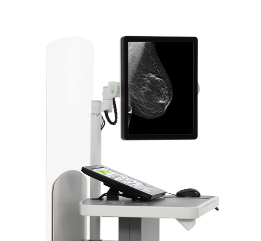
I-View™ Contrast Enhanced Imaging
Turn the invisible into the visible and detect cancers that can be hard to see on standard mammograms.
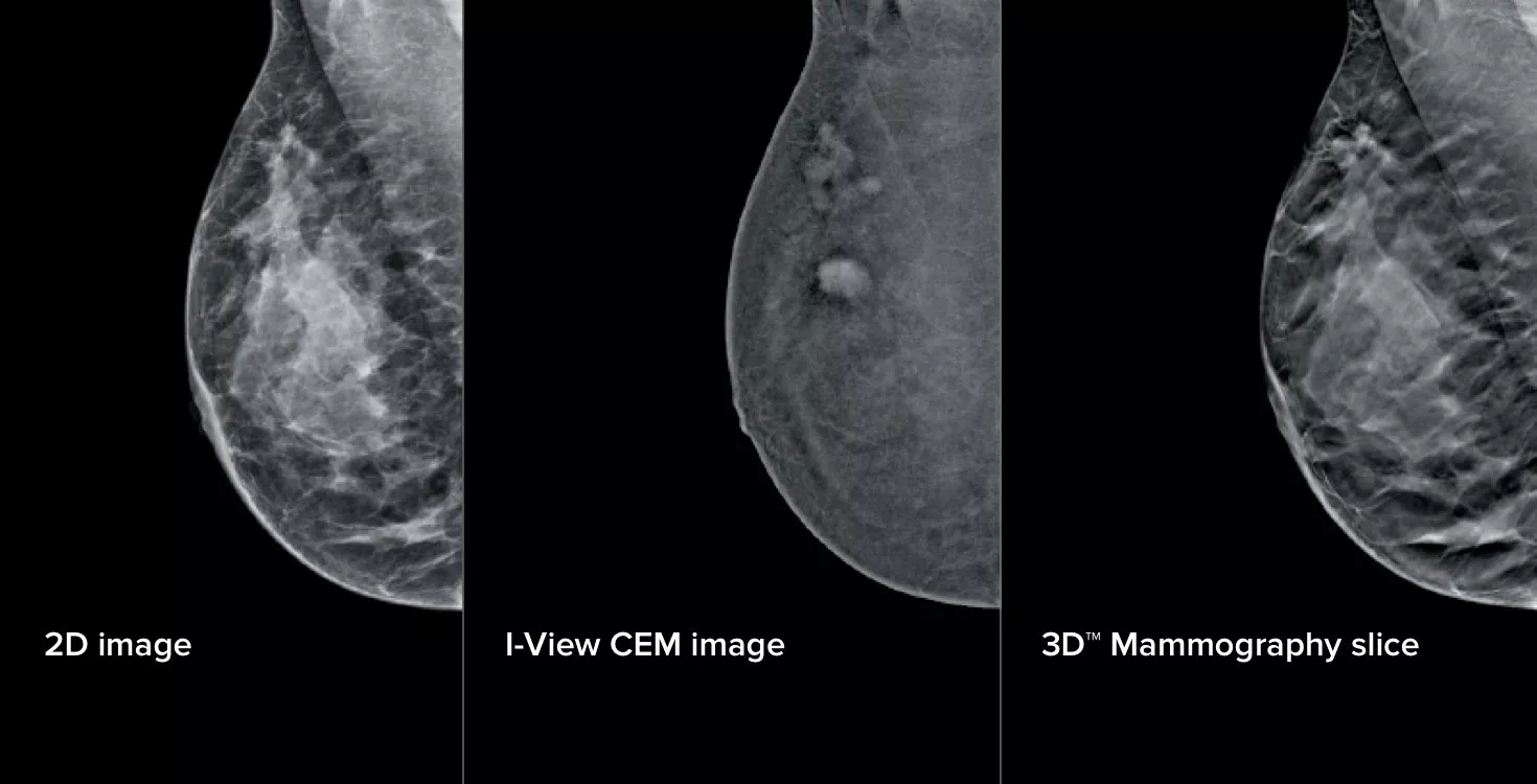
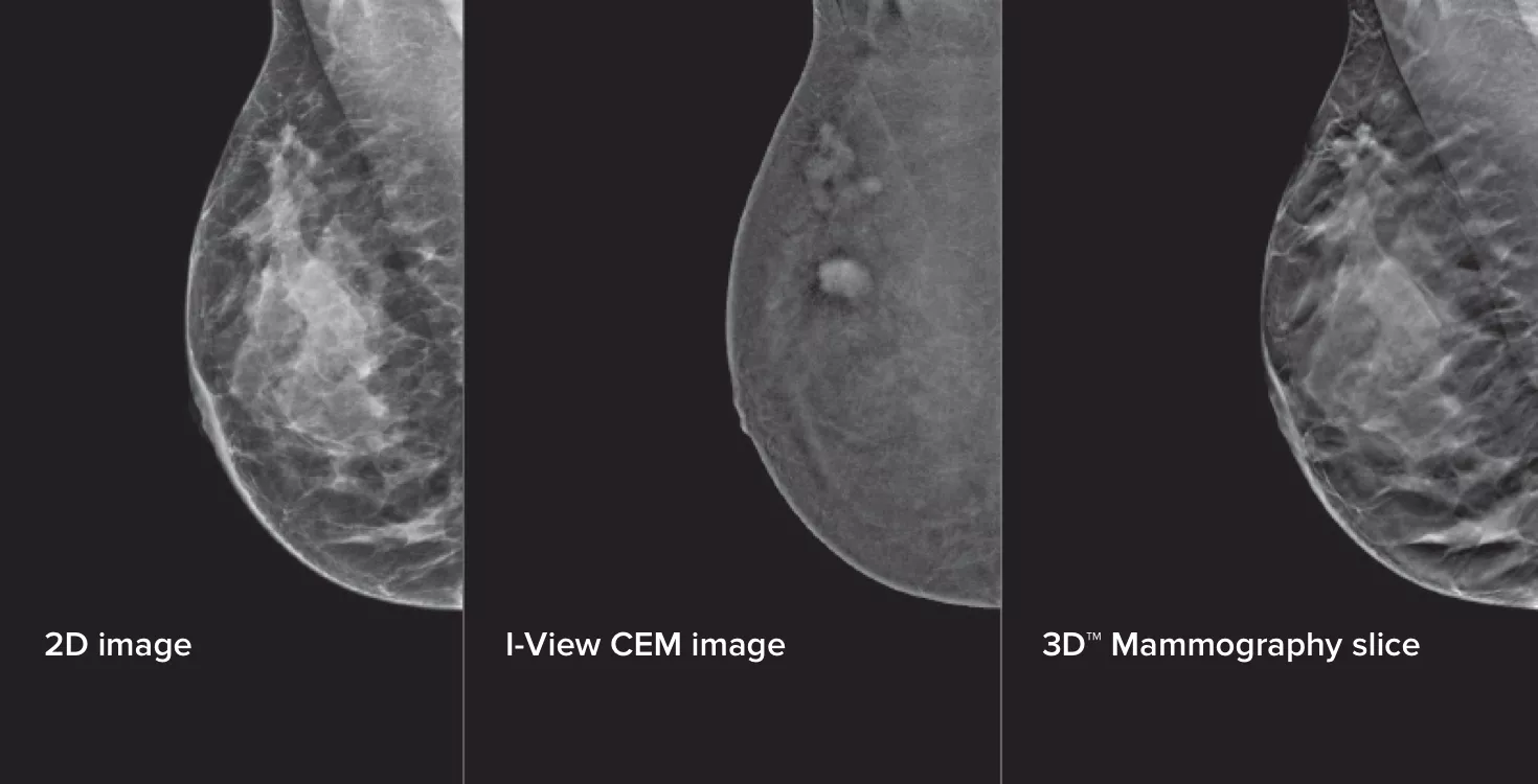
3-in-1 Contrast Enhanced Mammography
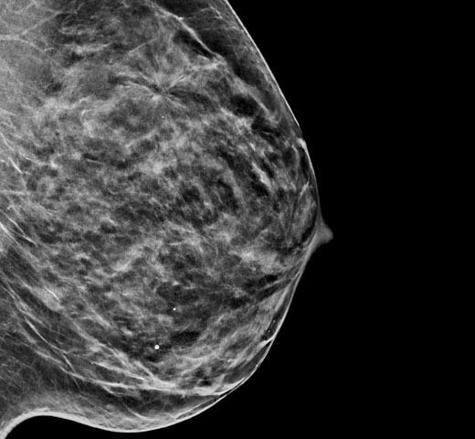
Contrast Enhanced Mammography (CEM), the imaging of a breast using iodinated contrast to reveal areas of increased blood supply within the breast, can help enhance suspicious lesions. The I-View software can combine the power of CEM with 2D and tomosynthesis images, all under one compression, providing anatomical and functional imaging in one exam.1
CEM is a valuable, compassionate and time-efficient breast diagnostic imaging solution. It compares favourably with breast MRI, offering similar sensitivity and higher specificity, with higher positive predictive value (PPV),2,3 making it a viable and cost-effective imaging modality.4
Sensitive, High Specificity & Cost-Effective
Comprehensive 3-in-1 Exam
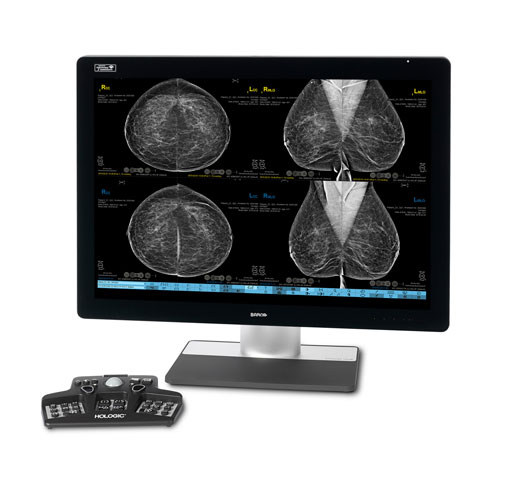
Accelerate your reading time with comprehensive imaging, using co-registered functional and morphological information.
Improved Patient Experience
Increasing the radiologist’s diagnostic confidence with high sensitivity and specificity imaging, helping to guide the clinical pathway from diagnosis to surgical management.
Efficient Clinical Workflow
The #1 adjunct diagnostic exam for women with dense breasts, and a quicker alternative to MRI.5
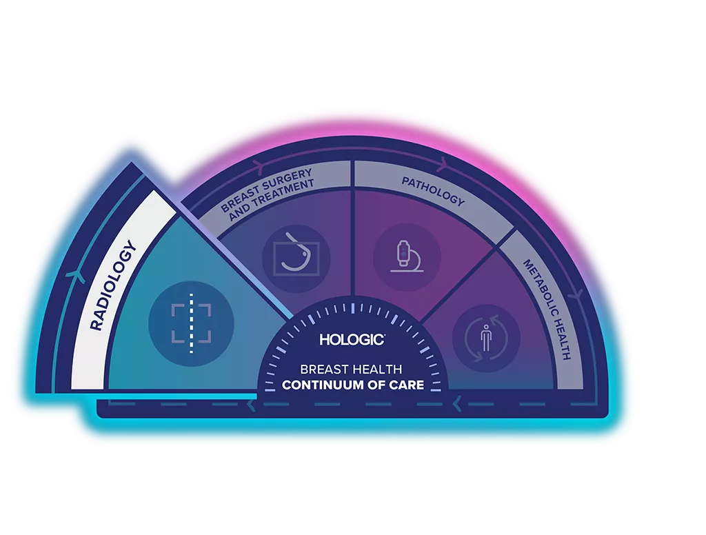
Unlock the Advantage of Time
The Breast Health Continuum of Care offers integrated solutions for clinical confidence, workflow efficiency and compassionate patient care. It gives more women, more time in better health.
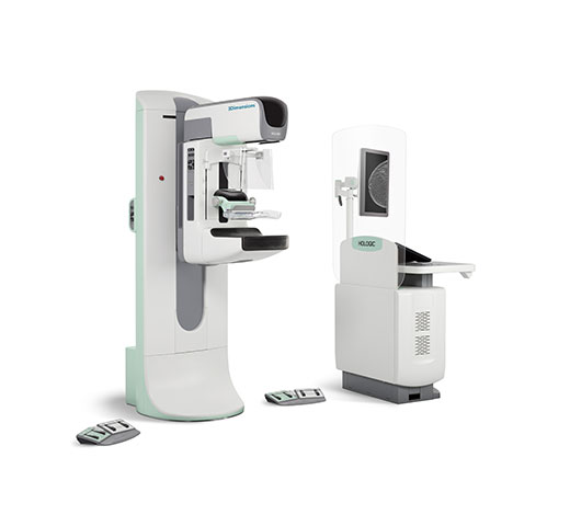
I-View Contrast Enhanced Imaging is part of the Hologic Screening and Diagnosis Solution.
Improve the Experience & Save Time
CEM mammography is quicker compared to MRI, increases patient compliance and improves their experience.6
8-20 min
imaging time
30 min
room block required
25%
of the cost of MRI6
79%
of patients prefer CEM over MRI7
3 Images from 1 Compression
This software captures both anatomical and functional information in a single exam by leveraging our ability to provide 2D, contrast and tomosynthesis images in just one compression.1
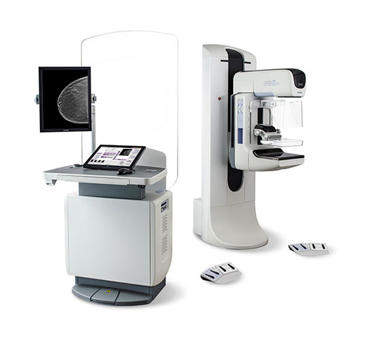
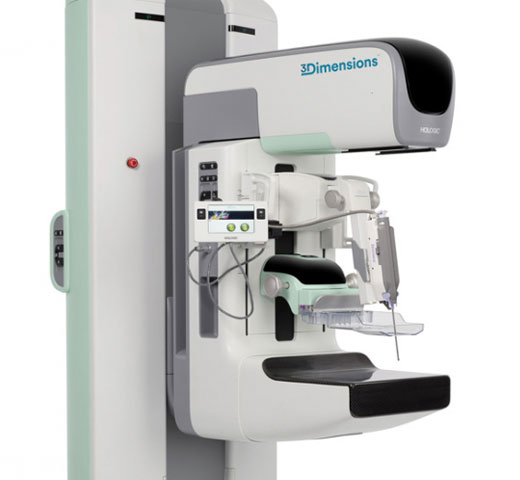
I-View CEM imaging is a simple upgrade to any Selenia® Dimensions®* and 3Dimensions™ system, giving breast imaging practices an efficient pathway to expanded diagnostic capabilities.
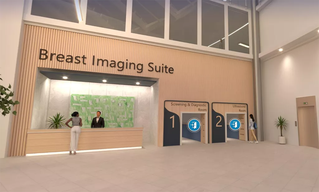
Visit Our Virtual Hospital
Browse our portfolio of Breast Health solutions in 3D. See how you can unlock the advantage of time across the entire Breast Continuum of Care.
Evidence. Insight. Collaboration.
Our education portal improves patient care through excellence in education, communication of clinical and scientific evidence, and partnerships with the healthcare community.
Insights
- Burhenne LJW., Wood SA., D'Orsi CJ., et al. Potential Contribution of Computer-aided Detection to the Sensitivity of Screening Mammography, Radiology 2000; 215:554-562;
- Chou C., Lewin JM., Chiang C., et al. Clinical Evaluation of Contrast-Enhanced Digital Mammography and Contrast Enhanced Tomosynthesis-Comparison to Contrast-Enhanced Breast MRI, Eur J Radiol. 2015 Dec; 84(12):2501-8. [Epub 2015 Oct 1].
- Li L., Roth R., Germaine P., et al. Contrast-enhanced spectral mammography (CESM) versus breast magnetic resonance imaging (MRI): A retrospective comparison in 66 breast lesions. Diagnostic and Interventional imaging Feb 2017.
- Xing D., Lv Y., Sun B., et al. Diagnostic Value of Contrast-Enhanced Spectral Mammography in Comparison to Magnetic Resonance Imaging in Breast Lesions. J Comput Assist Tomogr. Mar/Apr 2019.
- Li L., Roth R., Germaine P., et al, Contrast-enhanced spectral mammography (CESM) versus breast magnetic resonance imaging (MRI): A retrospective comparison in 66 breast lesions. Diagnostic and Interventional imaging Feb 2017.
- Breast MRI - Available at: https://www.radiologyinfo.org/en/info.cfm?pg=breastmr= (Accessed in Nov 2022)
- Phillips J., Miller MM., Mehta TS., et al, Contrast-enhanced spectral mammography (CESM) versus MRI in the high-risk screening setting: patient preferences and attitudes. Clin Imaging. 2017 Mar-Apr;42:193-197. doi: 10.1016


