Diagnose Breast Cancer with Confidence
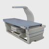
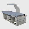
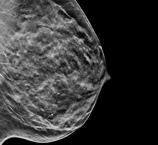
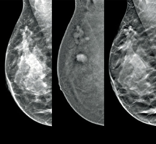
The Selenia Dimensions system delivers proven accuracy of our 3D Mammography exam to detect significantly more invasive breast cancers earlier and reduce call backs vs 2D alone.2-6,*
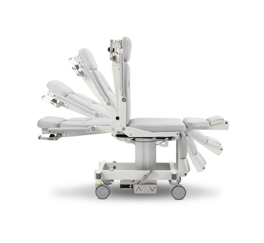
The system comes in two main configurations, to which you can add a range of options. This gives you and your department the flexibility to upscale the system with your needs. It's a choice already made by many departments, with over 15,000 systems** installed around the world.
Diagnose Challenging Patients with Greater Certainty
Reduce Recalls
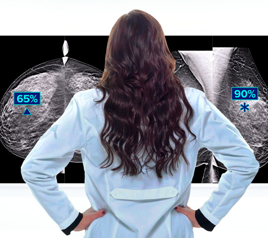
Detects up to 65% more invasive breast cancer, and reduces recalls by up to 40%, compared to 2D alone.2-4,*
Superior Accuracy
Superior accuracy for women with dense breasts compared to 2D alone.6
Faster Scans
Faster scans for greater comfort, a 3.7 second scan time for a 3D Mammography exam.7,†
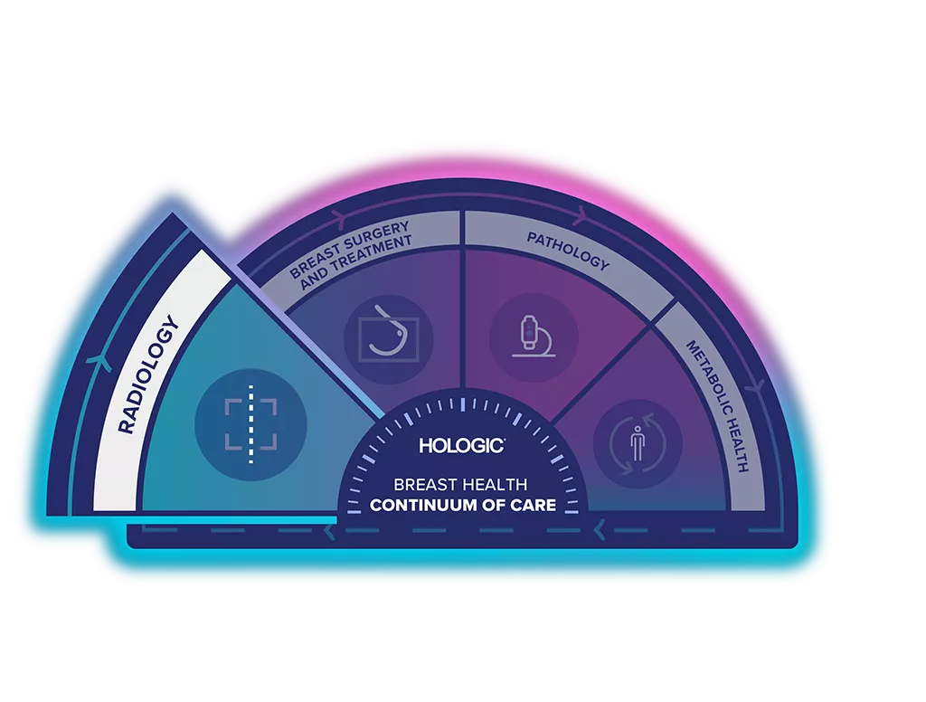
Unlock the Advantage of Time
The Breast Health Continuum of Care offers integrated solutions for clinical confidence, workflow efficiency and compassionate patient care. It gives more women, more time in better health.
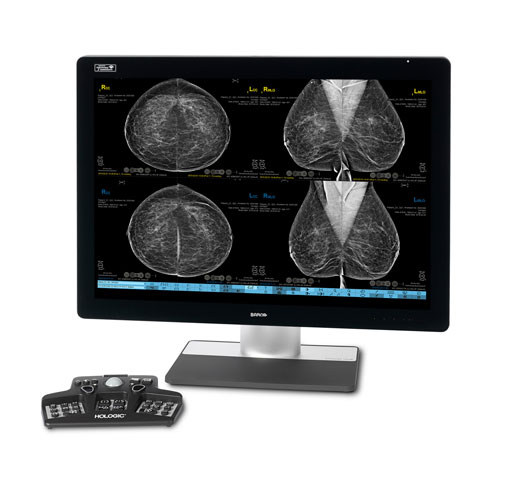
The Selenia Dimensions Digital Mammography System is part of the Hologic Screening & Diagnosis Solution.
Discover a Package that Works for You
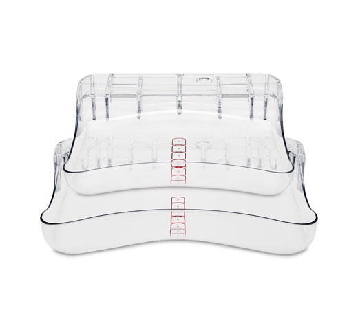
Hologic offers you two Selenia Dimensions mammography systems, each available in 2D, 3D imaging, and mobile packages. All packages are built around the way you work. They deliver the intelligent design, exceptional efficiency, and outstanding image quality needed to diagnose breast cancer with confidence. No matter which Selenia Dimensions package is right for you, you’ll be making an investment that pays, both now and in the future.
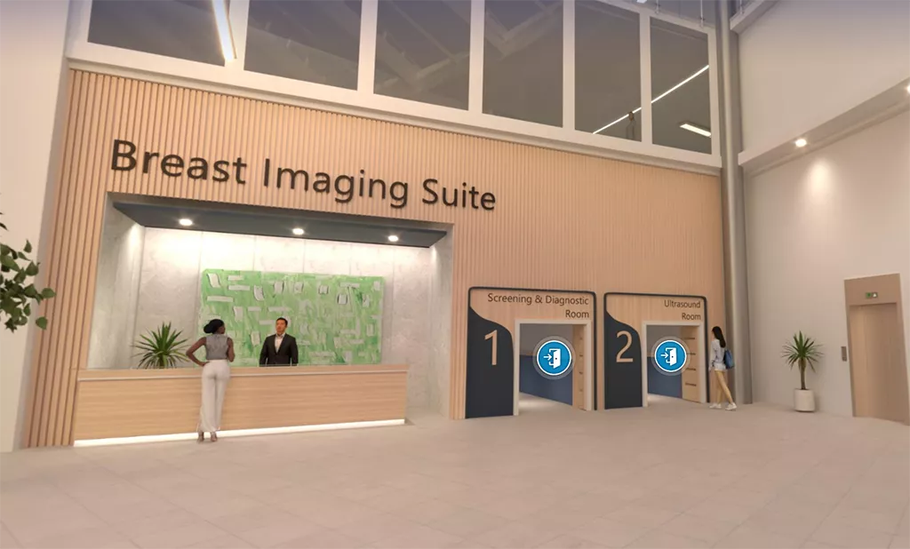
Visit Our Virtual Hospital
Browse our portfolio of Breast Health solutions in 3D. See how you can unlock the advantage of time across the entire Breast Continuum of Care.
Clinical Images
Video Gallery
Evidence. Insight. Collaboration.
Our education portal improves patient care through excellence in education, communication of clinical and scientific evidence, and partnerships with the healthcare community.
Insights
* Breast cancer screening using tomosynthesis in combination with digital mammography." JAMA 311.24 (2014): 2499-2507; a multi-site (13), non-randomized, historical control study of 454,000 screening mammograms investigating the initial impact the introduction of the Hologic Selenia® Dimensions ® on screening outcomes. Individual results may vary. The study found an average 41% (95% CI: 20-65%) increase and that 1.2 (95% CI: 0.8-1.6) additional invasive breast cancers per 1000 screening exams were found in women receiving combined 2D FFDM and 3D™ mammograms acquired with the Hologic 3D Mammography™ System versus women receiving 2D FFDM mammograms only
** Based on commercial data, October 2022
† Compared to other standard models
-
Tomosynthesis Bibliography: (PDF) 3D Mammography™ Bibliography - Hologic Education (Accessed Nov 2022)
-
Friedewald SM., Rafferty EA, Rose SL., et al. Breast cancer screening using tomosynthesis in combination with digital mammography. JAMA. 2014 Jun 25;311(24):2499-507.
-
Rose SL, Tidwell AL., Bujnoch LJ., et al. Implementation of breast tomosynthesis in a routine screening practice: an observational study. AJR AmJ Roentgenol. 2013;200(6):1401-1408
-
Haas BM., Kalra V., Geisel J., et al. Comparison of tomosynthesis plus digital mammography and digital mammography alone for breast cancer screening. Radiology 2013;269:694-700.
-
McDonald ES, Oustimov A, Weinstein SP, et al. Effectiveness of Digital Breast Tomosynthesis Compared With Digital Mammography: Outcomes Analysis From 3 Years of Breast Cancer Screening. JAMA Oncol. 2016 Jun 1;2(6):737-43.
-
Rafferty EA, Durand MA, Conant EF, et al. Breast Cancer Screening Using Tomosynthesis and Digital Mammography in Dense and Nondense Breasts. JAMA. 2016 Apr 26;315(16):1784-6. doi: 10.1001/jama.2016.1708. PMID: 27115381.
-
Rocha Garcia, A.M., Mera Fernandez, D. Breast tomosynthesis: State of the art. Radiologia. 2019;61(4):274-285.

